Upper Leg Tendon Anatomy : How To Remember The Muscles Of The Upper Thigh Youtube / Front view of knee after patellar tendon repair.. The primary sutures repair the torn tendon and the relaxing suture encompasses the repair and goes around the There are three on the back of the leg for plantar flexion, gastrocnemius, soleus, and plantaris. Eversion and plantarflexion of the foot. May 31, 2021 · gastrocnemius is a large muscle located in the posterior leg.posteriorly, is the most superficial of the muscles of the leg, and forms the bulk of the calf.it takes its name from the greek words γαστήρ (gaster) meaning stomach or belly, and κνήμη (kneme) meaning leg; Front view of knee after patellar tendon repair.
The combination of the two words means the "belly of the leg" or in other words the bulk of the calf. Front view of knee after patellar tendon repair. There's one muscle on the front of the leg for dorsiflexion, tibialis anterior. During an open surgery, an incision is made in the back of the leg and the achilles tendon is stitched together. Apr 01, 2018 · the fibres converge into a tendon, which descends into the foot, posterior to the lateral malleolus.
13 843 Best Leg Muscle Anatomy Images Stock Photos Vectors Adobe Stock from t4.ftcdn.net Eversion and plantarflexion of the foot. Quadriceps tendon patella patellar tendon figure 2. Its tendon is often called the freshman nerve because it is often misidentified by the freshman medical student Front view of knee after patellar tendon repair. There are three on the back of the leg for plantar flexion, gastrocnemius, soleus, and plantaris. Originating below and beneath the gastrocnemius is the soleus muscle, which extends your foot when your knee is bent. Also supports the lateral and transverse arches of the foot. Apr 23, 2019 · the plantaris is a small muscle with a long tendon, which can be mistaken for a nerve as it descends down the leg.
Front view of knee after patellar tendon repair.
Eversion and plantarflexion of the foot. Apr 23, 2019 · the plantaris is a small muscle with a long tendon, which can be mistaken for a nerve as it descends down the leg. In a complete or serious rupture the tendon of plantaris or another vestigial muscle is harvested and wrapped around the achilles tendon, increasing the strength of the repaired tendon. Apr 01, 2018 · the fibres converge into a tendon, which descends into the foot, posterior to the lateral malleolus. May 31, 2021 · gastrocnemius is a large muscle located in the posterior leg.posteriorly, is the most superficial of the muscles of the leg, and forms the bulk of the calf.it takes its name from the greek words γαστήρ (gaster) meaning stomach or belly, and κνήμη (kneme) meaning leg; Also supports the lateral and transverse arches of the foot. It serves to attach the plantaris, gastrocnemius (calf) and soleus muscles to the calcaneus (heel) bone. Its tendon is often called the freshman nerve because it is often misidentified by the freshman medical student Dorsum of the calcaneus medial to the calcaneal tendon: Plantaris has a long slender tendon that is equivalent to the tendon of the palmaris longus m. The muscle descends medially, condensing into a tendon that runs down the leg, between the gastrocnemius and soleus. Front view of knee after patellar tendon repair. Originating below and beneath the gastrocnemius is the soleus muscle, which extends your foot when your knee is bent.
Originates from the lateral supracondylar line of the femur. The tendon crosses under the foot, and attaches to the bones on the medial side, namely the medial cuneiform and base of metatarsal i. The achilles tendon or heel cord, also known as the calcaneal tendon, is a tendon at the back of the lower leg, and is the thickest in the human body. It serves to attach the plantaris, gastrocnemius (calf) and soleus muscles to the calcaneus (heel) bone. Its tendon is often called the freshman nerve because it is often misidentified by the freshman medical student
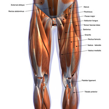
Upper Legs Muscles Anatomy 3d Render Stock Photo Picture And Low Budget Royalty Free Image Pic Esy 030353373 Agefotostock from previews.agefotostock.com There's one muscle on the front of the leg for dorsiflexion, tibialis anterior. It is absent in 10% of people. Quadriceps tendon patella patellar tendon figure 2. There are three on the back of the leg for plantar flexion, gastrocnemius, soleus, and plantaris. The muscle descends medially, condensing into a tendon that runs down the leg, between the gastrocnemius and soleus. Dorsum of the calcaneus medial to the calcaneal tendon: During an open surgery, an incision is made in the back of the leg and the achilles tendon is stitched together. Apr 01, 2018 · the fibres converge into a tendon, which descends into the foot, posterior to the lateral malleolus.
Front view of knee after patellar tendon repair. Apr 23, 2019 · the plantaris is a small muscle with a long tendon, which can be mistaken for a nerve as it descends down the leg. It is absent in 10% of people. It serves to attach the plantaris, gastrocnemius (calf) and soleus muscles to the calcaneus (heel) bone. The tendon crosses under the foot, and attaches to the bones on the medial side, namely the medial cuneiform and base of metatarsal i. Dorsum of the calcaneus medial to the calcaneal tendon: Apr 01, 2018 · the fibres converge into a tendon, which descends into the foot, posterior to the lateral malleolus. Its tendon is often called the freshman nerve because it is often misidentified by the freshman medical student There are three on the back of the leg for plantar flexion, gastrocnemius, soleus, and plantaris. Plantaris has a long slender tendon that is equivalent to the tendon of the palmaris longus m. Quadriceps tendon patella patellar tendon figure 2. In a complete or serious rupture the tendon of plantaris or another vestigial muscle is harvested and wrapped around the achilles tendon, increasing the strength of the repaired tendon. Tibialis anterior arises from the lateral surface of the upper tibia, and from the interosseous membrane.
Tibialis anterior arises from the lateral surface of the upper tibia, and from the interosseous membrane. Apr 23, 2019 · the plantaris is a small muscle with a long tendon, which can be mistaken for a nerve as it descends down the leg. There are three on the back of the leg for plantar flexion, gastrocnemius, soleus, and plantaris. Originates from the lateral supracondylar line of the femur. Quadriceps tendon patella patellar tendon figure 2.
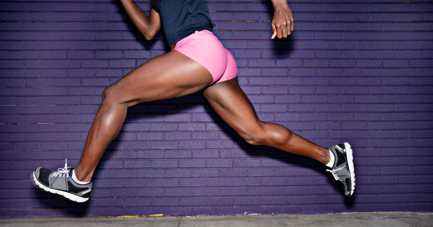
Anatomy Of Your Leg Muscles Plus How Genetics And Exercise Can Change Your Leg Muscles Shape from imagesvc.meredithcorp.io During an open surgery, an incision is made in the back of the leg and the achilles tendon is stitched together. It is absent in 10% of people. Also supports the lateral and transverse arches of the foot. Its tendon is often called the freshman nerve because it is often misidentified by the freshman medical student Plantaris has a long slender tendon that is equivalent to the tendon of the palmaris longus m. Eversion and plantarflexion of the foot. Originates from the lateral supracondylar line of the femur. It serves to attach the plantaris, gastrocnemius (calf) and soleus muscles to the calcaneus (heel) bone.
In a complete or serious rupture the tendon of plantaris or another vestigial muscle is harvested and wrapped around the achilles tendon, increasing the strength of the repaired tendon.
Eversion and plantarflexion of the foot. The muscle descends medially, condensing into a tendon that runs down the leg, between the gastrocnemius and soleus. Originating below and beneath the gastrocnemius is the soleus muscle, which extends your foot when your knee is bent. Originates from the lateral supracondylar line of the femur. It serves to attach the plantaris, gastrocnemius (calf) and soleus muscles to the calcaneus (heel) bone. The combination of the two words means the "belly of the leg" or in other words the bulk of the calf. Apr 23, 2019 · the plantaris is a small muscle with a long tendon, which can be mistaken for a nerve as it descends down the leg. The primary sutures repair the torn tendon and the relaxing suture encompasses the repair and goes around the Also supports the lateral and transverse arches of the foot. During an open surgery, an incision is made in the back of the leg and the achilles tendon is stitched together. Quadriceps tendon patella patellar tendon figure 2. Dorsum of the calcaneus medial to the calcaneal tendon: Apr 01, 2018 · the fibres converge into a tendon, which descends into the foot, posterior to the lateral malleolus.


Posting Komentar
0 Komentar